June 2006

Mass at the upperpole of the left kidney

Clinical information

Incidental finding in a patient examined because of a diverticulitis

Ultrasound Images & Clips

77 years old female, Incidental finding in a patient examined because of a diverticulitis

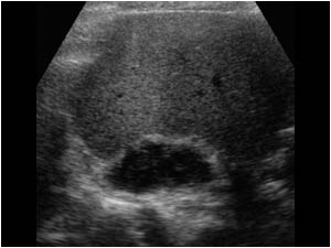
Transverse image of a hypoechoic mass at the upperpole of the left kidney.
Longitudinal image of the hypoechoic mass at the upperpole of the left kidney invading the kidney.
Transverse image of the hypoechoic mass at the upperpole of the left kidney invading the kidney.
Transverse image of the same patient showing a smaller hypoechoic mass at the upperpole of the right kidney.
 Longitudinal image of the smaller hypoechoic mass at the upperpole of the right kidney.
Longitudinal image of the smaller hypoechoic mass at the upperpole of the right kidney.
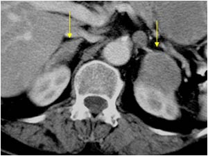
CT image showing the two masses a the upper poles of the kidneys.
CT image at a lower level showing the two masses at the upper poles of the kidneys. The left one invading the kidney.
CT image of a CT guided biopsy showing the needle just below the pleural sinus.
 CT image of a CT guided biopsy showing the needle in the mass in the upperpole of the left kidney.

The biopsy proved the mass to be a metastasis of a lung carcinoma.
CT image of a CT guided biopsy showing the needle in the mass in the upperpole of the left kidney. The biopsy proved the mass to be a metastasis of a lung carcinoma.
An X ray of the chest shows a small lung carcinoma confirmed with bronchoscopy.

Conclusion

Diagnosis: Bilateral adrenal metastases of a small lung carcinoma. The largest adrenal metastasis invades the left kidney so that it could be mistaken for a renal tumor.

Details

  • Sex: Female
  • Age: 77

Created with

  • Fujifilm Ultrasound System