July 2006

Abnormal Scrotum

Clinical information

Diffuse moderately painful swelling of the scrotum and erythema
Ultrasound Images & Clips

6 year old boy, Diffuse moderately painful swelling of the scrotum and erythema
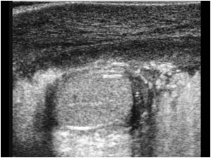
Longitudinal image of the right scrotum with a normal testicle and peritesticular structures. No hydrocele. The right scrotal wall is markedly thickened.
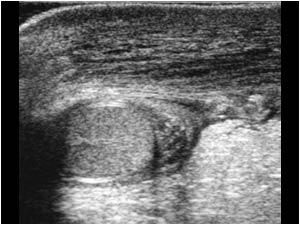
Longitudinal image of the left scrotum also with a normal testicle and peritesticular structures. No hydrocele. The left scrotal wall is markedly thickened.
Transverse image of the right testicle with the same findings.
Transverse image of the left testicle also with the same findings.
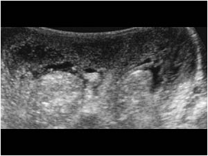
AP image of the swollen scrotum.
 Lateral image showing the scrotal swelling and erythema.
Lateral image showing the scrotal swelling and erythema.
Convex transverse image showing an edematous scrotal wall and normal testicles and peritesticular structures.
Longitudinal image of the right testicle showing normal vascularity of the testis and peritesticular structures.
 Longitudunal image of the left testicle showing normal vascularity of the testis and peritesticular structures.
Longitudunal image of the left testicle showing normal vascularity of the testis and peritesticular structures.
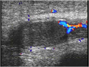
 Color doppler image of the scrotal wall showing markedly increased vascularity.
Color doppler image of the scrotal wall showing markedly increased vascularity.

Conclusion

Because the testicle and peritesticular structures were normal a testicular torsion could be excluded. Also an epididymitis and torsion of an appendix testis could be excluded. Because of the massive edematous swelling of the scrotal wall with markedly increased vascularity the findings are consistent with an idiopathic scrotal edema. This is a self-limiting acute scrotal edema and erythema that resolves without sequela. Idiopathic scrotal edema occurs mostly in children under 10 years of age. Although it is usualy unilateral bilateral cases have been reported.

References
Acute idiopathic scrotal edema in children revisited. Klin B, Lotan G, Efrati Y, Zlotkevich L, Strauss S.
J Pediatr Surg. 2002 Aug;37(8):1200-2.

Acute idiopathic scrotal oedema: four cases and a short review. van Langen AM, Gal S, Hulsmann AR, De Nef JJ
Eur J Pediatr. 2001 Jul;160(7):455-6.

Details

  • Sex: Male
  • Age: 6

Created with

  • Fujifilm Ultrasound System