November 2006

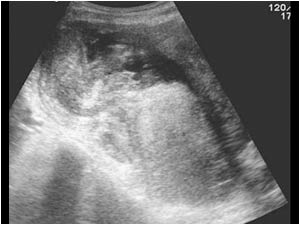
Partly cystic and partly solid mass of more than 10 cm in the right lower abdomen. During a laparoscopy a few months earlier normal ovaries were found.

Clinical information

Persistent pain in the right lower abdomen, raised sedimentation rate and a palpable mass.
Ultrasound Images & Clips

35 year old female, Persistent pain in the right lower abdomen, raised sedimentation rate and a palpable mass.

Longitudinal image of the partly cystic and partly solid mass of more than 10 cm in the right lower abdomen.
Transverse image of the mass.
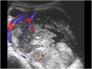
Longitudinal detail image of the mass showing vascularity in the periphery of the lesion, but not in the central part.
Transverse color Doppler image showing peripheral vascularity
CT image of the same lesion showing a large mainly cystic mass.
Partly cystic and partly solid mass of more than 10 cm in the right lower abdomen. During a laparoscopy a few months earlier normal ovaries were found.
Two CT scan images of the same patient made a few month earlier when the patient was admitted under suspicion of having a pelvic inflammatory disease or an appendicitis . The images show a smaller irregular mass with cystic areas. A laparoscopy was perfor

Conclusion

The symptoms diminished, but a repeated CT scan still showed the mass. The patient was released from the hospital but returned after a few months because she had persistent pain.

Because of the cystic nature of the mass and the inflammatory symptoms the first impression was that the mass could be an appendiceal abscess. Although the patient was ill, there was doubt about the nature of the mass. Instead of doing a percutaneous drainage as requested by the surgeon, it was decided to biopsy the solid part of the mass under ultrasound guidance first to exclude a malignancy. Unfortunately for the patient the cystic mass proved to be a mucinous carcinoma. The patient was referred to a special cancer centre were she was successfully operated.

Appendiceal carcinoma is rare. Mucinous carcinoma is the most common form of appendiceal carcinoma. Usually the diagnosis is made by laparoscopy.

Differentiation from inflammatory disease, a mucocele of the appendix or ovarian tumors can be very difficult.

References
McGory ML, Maggard MA, Kang H, O'Connell JB, Ko CY.
Malignancies of the appendix: beyond case series reports.
Dis Colon Rectum. 2005 Dec;48(12):2264-71.

Details

  • Sex: Female
  • Age: 35

Created with

  • Fujifilm Ultrasound System