December 2006

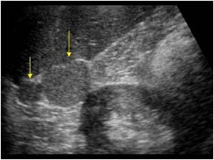
Two circumscribed lesions cranial of the right kidney.

Clinical information

Incidental finding during examination of the kidneys. Abdominal surgery during infancy after trauma decades before.
Ultrasound Images & Clips

44 year old male, Incidental finding during examination of the kidneys. Abdominal surgery during infancy after trauma decades before.
Two circumscribed lesions cranial of the right kidney.
Image of the upperpole of the right kidney with two circumscribed lesions between the kidney and right liver lobe.
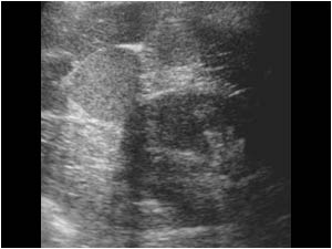
Transverse image of one of the lesions caudal of the right liver lobe.
Transverse image of the right kidney and one of the lesions. The lesion is situated lateral to the kidney and is not situated in the area of the right adrenal.
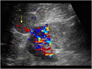
 Color doppler image of the kidney and one of the lesions shows vascularization of the mass.

During examination no spleen could be found. Patient had undergone a splenectomy as a child after an abdominal trauma with a splenic rupture.

This informati
Color doppler image of the kidney and one of the lesions shows vascularization of the mass. During examination no spleen could be found. Patient had undergone a splenectomy as a child after an abdominal trauma with a splenic rupture. This informati
The lesions look like splenic tissue as can be seen in accessory spleens, as is shown in this normal patient with two accessory spleens.
Image of the left flank in another patient with an accessory spleen after splenectomy.

Conclusion

In order to prove that the patient really had a splenosis with two masses with ectopic splenic tissue in the right flank after a splenic rupture, an isotope study was performed in a university hospital elswhere. The isotope study showed two small structures with functional splenic tissue at the site of the two lesions

Diagnosis Splenosis with ectopic splenic tissue after a splenic rupture.

References
Khosravi MR, Margulies DR, Alsabeh R, Nissen N, Phillips EH, Morgenstern L. Consider the diagnosis of splenosis for soft tissue masses long after any splenic injury.
Am Surg. 2004 Nov;70(11):967-70.

Wedemeyer J, Gratz KF, Soudah B, Rosenthal H, Strassburg C, Splenosis--important differential diagnosis in splenectomized patients presenting with abdominal masses of unknown origin]
Z Gastroenterol. 2005 Nov;43(11):1225-9.

Details

  • Sex: Male
  • Age: 44

Created with

  • Fujifilm Ultrasound System