January 2008
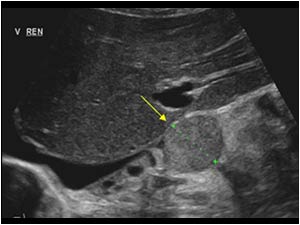
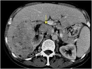
Abnormal aspect of the inferior vena cava
[{"listLocation":"abdomen-and-retroperitoneum","icon":"001-abdomen-white.svg","header":"Abdomen and retroperitoneum","id":63},{"listLocation":"urinary-tract-and-male-reproductive-system","icon":"002-urinary-tract-white.svg","header":"Urinary Tract and male reproductive system","id":64},{"listLocation":"gynaecology","icon":"003-gynaecology-white.svg","header":"Gynaecology","id":65},{"listLocation":"head-and-neck","icon":"004-head-neck-white.svg","header":"Head and Neck","id":66},{"listLocation":"breast-and-axilla","icon":"005-breast-white.svg","header":"Breast and Axilla","id":67},{"listLocation":"musculo-skeletal-joints-and-tendons","icon":"006-msk-joints-white.svg","header":"Musculoskeletal Joints and Tendons","id":68},{"listLocation":"musculo-skeletal-bone-muscle-nerves-and-other-soft-tissues","icon":"007-msk-bones-white.svg","header":"Musculoskeletal, bone, muscle, nerves and other soft tissues","id":69},{"listLocation":"thorax","icon":"008-thorax-white.svg","header":"Thorax","id":70},{"listLocation":"pediatrics","icon":"009-pediatrics-white.svg","header":"Pediatrics","id":71},{"listLocation":"peripheral-vessels","icon":"010-peripheral-vessels-white.svg","header":"Peripheral vessels","id":72}]

