January 2010

Anechoic structure in the abdominal wall in the umbilical region

Clinical information

2 male patients age 64 and 71
Both patients have a palpable lesion in the abdominal wall near the umbilicus. One shows a slight blue discoloration. An ultrasound examination was requested to confirm an umbilical hernia.
Ultrasound Images & Clips

2 male patients age 64 and 71, Both patients have a palpable lesion in the abdominal wall near the umbilicus. One shows a slight blue discoloration. An ultrasound examination was requested to confirm an umbilical hernia.
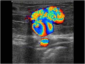
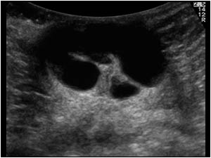
Transverse image of the lesion in patient 1
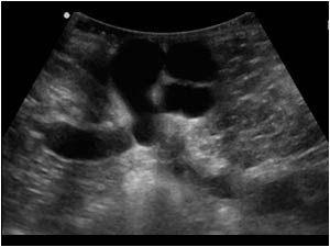
Transverse image of the lesion in patient 2.
Can you guess what the lesion is and what is the underlying problem?
Transverse image of the lesion in patient 2. Can you guess what the lesion is and what is the underlying problem?
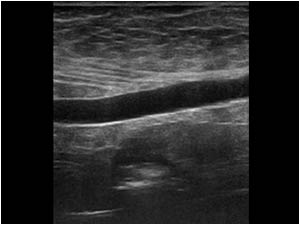
Longitudinal image of the lesion in patient 2.
You have probably noticed that both lesions are no umbilical hernias, but look like dilatated vascular structures.
Longitudinal image of the lesion in patient 2. You have probably noticed that both lesions are no umbilical hernias, but look like dilatated vascular structures.
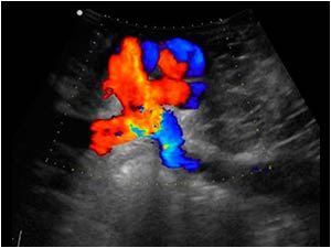
Longitudinal color Doppler image of the lesion of patient 2. There is flow and there is a vessel entering and leaving the lesion.
Transverse color Doppler image of the lesion of patient 1 also showing flow.
Both lesions represent a herniating paraumbilical vein varix.
Did you notice the vessel directly underneath the abdominal wall?
Transverse color Doppler image of the lesion of patient 1 also showing flow. Both lesions represent a herniating paraumbilical vein varix. Did you notice the vessel directly underneath the abdominal wall?
The Doppler spectrum confirms the venous nature of the lesion in patient 1.
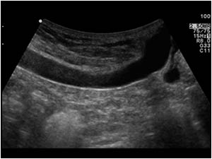
Longitudinal image of the vein underneath the abdominal wall of patient 1. Can you guess where the vein is coming from?
Longitudinal image of the upper abdomen of patient 2. There is also a vein running underneath the abdominal wall.
Longitudinal color Doppler image of patient 2. The vein can be followed all the way to the liver.
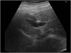
Transverse image of patient 2 showing the vein in relation to the liver.
In both patients the venous structure in the upper abdomen represents a recanalized umbilical vein or a widened paraumbilical vein.
A recanalized umbilical or a widened paraumbilic
Transverse image of patient 2 showing the vein in relation to the liver. In both patients the venous structure in the upper abdomen represents a recanalized umbilical vein or a widened paraumbilical vein. A recanalized umbilical or a widened paraumbilic
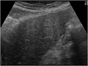
Longitudinal image of the left liver lobe of patient 2 showing a hyperechoic liver with an irregular contour.
Longitudinal image of the right liver lobe of patient 1 showing the same features.
Both patients have a cirrhotic liver.
Longitudinal image of the right liver lobe of patient 1 showing the same features. Both patients have a cirrhotic liver.
Anechoic structure in the abdominal wall in the umbilical region

Conclusion

In patients with liver cirrhosis it is important to look for other signs of portal hypertension. Both patients also had a splenomegaly as shown here on this longitudinal image of patient 1 showing an 18 cm large spleen.

Details

  • Sex: Male
  • Age: 64

Created with

  • Fujifilm Ultrasound System