November 2014

Oval lobulated partly well defined and partly ill defined mass that is there for several years but not biopsied

Clinical information

Has a slow-growing breast mass that is there since 2003. Patient was examined in 2003 but the lesion looked benign and was not biopsied. No images from 2003 are available. She returned in 2013.
Ultrasound Images & Clips

Oval lobulated mass that is there for several years but not biopsied

Oval mass that is for a part well defined and for a part not well defined
Detail image of the well defined part of the lesion
Transverse image of the well defined part
Detail of the nor so well defined part
The lesion is several cm's large
Mammography
Mammography
Mammography
Mammography
Image of an oval mass taken in 2008
Transverse image from the mass in 2008 Biopsy showed a fibroadenona
In march 2012 the lesion had grown rather fast
Transverse image of the mass
The mass is vascularized
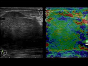
An elastogram showed a soft mass. Although the lesion has benign characteristics a repeat 14 G biopsy was performed and we asked the pathologist if the lesion could be a phylloides tumor
Again the result was fibroadenoma and half a year later the lesion had grown
The lesion is still vascularized
At the end of 2013 the lesion was so big thatwe had to use the extended field of view to get the mass in one image
Mammography in 2008
Mammography in 2008
Mammography in 2012
Mammography in 2012

Conclusion

Although the lesion of the first patient was according to the patient there for 10 years the lesion proved to be a malignant phyllodes tumor. No metastases were found. The second patient had a very fast growing phyllodes tumor that at the time of operation had grown to 14 cm. At operation there proved to be a small localized area of DCIS grade 2. The rest of the lesion, however, was still benign. The pathologist told us that differentiation between a fibroadenoma and a benign phyllodes tumor can be very difficult no matter the size of the biopsy needle.

Details

  • Sex: Female
  • Age: 58

Created with

  • Fujifilm Ultrasound System