April 2008

Abnormal aspect of the intrahepatic portal veins

Clinical information

Weight loss and abnormal liver function tests
Ultrasound Images & Clips

71 year old male, Weight loss and abnormal liver function tests
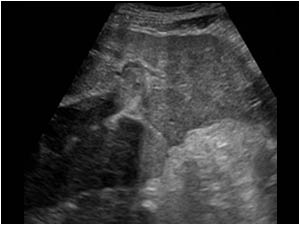
Transverse image of the left portal vein with an intraluminal structure
Detail of the liver contour showing an irregular liver contour
Another image of the liver showing an intraluminal structure in the portal vein and an irregular liver contour
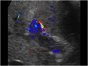
Detail of the portal vein shows no normal intraluminal flow, but color doppler signals suggestive of flow in the intraluminal mass
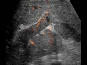
e- flow image of the portal vein also suggestive of flow in the intraluminal portal mass.

The rest of the liver parenchyma was diffuse inhomogeneous, but no circumscribed tumor could be found. A CT scan was recommended.
e- flow image of the portal vein also suggestive of flow in the intraluminal portal mass. The rest of the liver parenchyma was diffuse inhomogeneous, but no circumscribed tumor could be found. A CT scan was recommended.
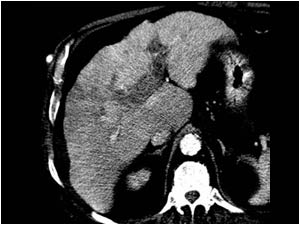
CT image of the same patient arterial phase
CT image of the same patient portal phase

The CT scan confirmed the irregular inhomogeneous aspect of the liver, but was not conclusive about the presence of a tumor or tumor vascularity in the portal veins.
Because the initial ultrasound examination
CT image of the same patient portal phase The CT scan confirmed the irregular inhomogeneous aspect of the liver, but was not conclusive about the presence of a tumor or tumor vascularity in the portal veins. Because the initial ultrasound examination
The cineloop clearly shows contrast enhancement in the intraluminal mass very suggestive of an extensive tumor thrombus in the portal veins. A biopsy to confirm the diagnosis was discussed but not performed.

Conclusion

The patient's condition deteriorated rapidly and he died a few weeks later. An autopsy was performed.
At autopsy a cirrhotic liver was found. In the portal veins a diffuse growing hepatocellular carcinoma was found.

References            
Rossi S, Rosa L, Ravetta V, Cascina A, Quaretti P, Azzaretti A, Scagnelli P, Tinelli C, Dionigi P, Calliada F. Contrast-enhanced versus conventional and color Doppler sonography for the detection of thrombosis of the portal and hepatic venous systems.
AJR Am J Roentgenol. 2006 Mar;186(3):763-73.

Tarantino L, Francica G, Sordelli I, Esposito F, Giorgio A, Sorrentino P, de Stefano G, Di Sarno A, Ferraioli G, Sperlongano P. Diagnosis of benign and malignant portal vein thrombosis in cirrhotic patients with hepatocellular carcinoma: color Doppler US, contrast-enhanced US, and fine-needle biopsyAbdom Imaging. 2006 Sep-Oct;31(5):537-44.

Details

  • Sex: Male
  • Age: 71

Created with

  • Fujifilm Ultrasound System