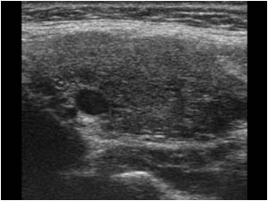
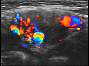
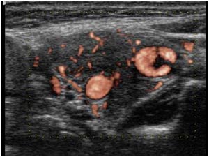
Painless palpable mass at the carotid artery bifurcation


The most common cause of a hypoechoic mass along the carotid arteries is an enlarged lymph node. The aspect of the lesion between the carotid arteries with encasement of the vessels and the irregular hypervascularity are atypical for a pathological lymph node.
In this case the mass represents a carotid body tumor.
This type of tumor was first described in 1762 by Haller and called a glomus tumor because of the glomus like structure of the tumor. Later the tumor was also named a chemodectoma or a paraganglioma.
Glomus tumors of the head and neck paraganglia are part of the extra-adrenal neuroendocrine system
Carotid body tumors usually present as a painless mass in the neck and tend to be asymptomatic. Because of its close relation to the carotid arteries they can pulsate. Sometimes a bruit can be heard over the mass.
The following features are commonly seen with ultrasound:
- A well-defined solid mass in the neck that may be unilateral or bilateral.
- The mass is usually hypoechoic. Anechoic tubular channels representing small vessels may be seen within the mass.
- The mass is found in close proximity to the carotid bifurcation often with widening of the bifurcation
- Color Doppler demonstrates significant internal vascularity within the mass The feeders arise from the external carotid artery, although the internal carotid and the vertebral artery may also supply the carotid body tumor.
-On spectral analysis, low resistance flow may be detected within the mass
References
Alkadhi H, et al. Evaluation of topography and vascularization of cervical paragangliomas by magnetic resonance imaging and color duplex sonography. Neuroradiology. 2002 Jan; 44(1):83-90.
Arslan H, Unal O, Kutluhan A, Sakarya ME. Power Doppler scanning in the diagnosis of carotid body tumors. J Ultrasound Med. 2000 Jun; 19(6):367-70.