January 2011

Normal retrocecal appendix????????

Clinical information

Pain in the right lower abdomen. Slightly raised infection parameters
Ultrasound Images & Clips

Normal retrocecal appendix????????

There is a normal terminal ileum.
The appendix is not in its most common position
There is a normal terminal ileum. The appendix is not in its most common position
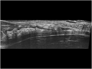
A normal looking appendix could be found in a retrocecal position
Longitudinal image of the normal looking appendix in a retrocecal position
CT scan of the same patient shows a normal appendix in a retrocecal position
Longitudinal image of the right kidney with a small amount of perirenal fluid
Transverse image of the right kidney with perirenal effusion
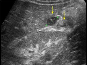
There was also a small effusion at the undersurface of the right liver lobe. What was not noticed was a small round structure next to this effusion
The folowing day the examination was repeated because the CRP had increased to 300
Again there is a rounded structure next to the effusion
The rounded structure was an inflamed tip of a very long retrocecal appendix. The largest part of the appendix still remained normal
videoclip
Videoclip of the same patient in longitudinal direction
CT scan of the same patient
The following case shows that the diagnosis of a focal appendicitis of the tip is not always easy
Detail of the tip of the appendix with vascularity
Transverse image of the tip with color doppler. The diameter is nearly 6 mm. which is within normal limits
Transverse image of the proximal appendix which measures 4,3 mm.

Conclusion

In the first case only the proximal part of the appendix was visualized in the first examinations. The inflamed tip was initially missed leading to a small perirenal effusion and an effusion at the lowerpole of the liver. A retrocecal appendix can be difficult to examine with ultrasound becuse of overlying bowel gas. When it is very long, a focal abnormality can easily be missed.
The second case illustrates that a focal thickening of the appendix doesn't always mean that there is an appendicitis. Although the rounded shape of the tip and the vasculatity were suspicious for inflammation, in this case it was still fysiological. The diameter was still within normal limits and there were no secundary signs of inflammation

References            
Optimizing US examination to detect the normal and abnormal appendix in children.
Peletti AB, Baldisserotto M.
Pediatr Radiol. 2006 Nov;36(11):1171-6.

US features of the normal appendix and surrounding area in children.
Wiersma F, Srámek A, Holscher HC.
Radiology. 2005 Jun;235(3):1018-22.

Details

  • Sex: Male
  • Age: 14

Created with

  • Fujifilm Ultrasound System