July 2019

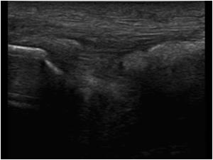
This patient has an abnormal patellar tendon. What could be the cause?

Clinical information

This patient has an abnormal patellar tendon. What could be the cause?

A) CPPD
B) Gout
C) R.A.
D) Tendinosis

Ultrasound Images & Clips

Patient with an abnormal patellar tendon

This patient has an abnormal patellar tendon.
What could be the cause?
A. CPPD
B. Gout
C. R.A.
D. Tendinosis
This patient has an abnormal patellar tendon. What could be the cause? A. CPPD B. Gout C. R.A. D. Tendinosis
Another longitudinal image of the abnormal patellar tendon
The calcifications cause acoustic shadowing
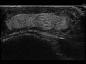
Transverse image of the patellar tendon
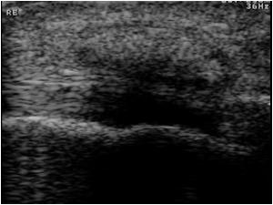
Another image of the same patient.
The patient was known to have long standing gout
Another image of the same patient. The patient was known to have long standing gout
Gout can  present itself in many joints. This is the olecranon bursa filled with echogenic material in a patient with gout
Gout can present itself in many joints. This is the olecranon bursa filled with echogenic material in a patient with gout
This is a transverse image of the olecranon fossa of a patient with gout
This is a longitudinal image of the same patient with synovial swelling and a bony erosion
This is an image of the superficial calcaneal bursa filled with echogenic material in a patient with gout
Detail image of cartilage calcifications in a patient with gout
This same patient also has synovial thickening with tiny calcifications
Transverse image of the same patient with synovial thickening with tiny calcifications
This is an image of the first metatarsophalangeal joint in a patient with gout with  synovial thickening with tiny calcifications
This is an image of the first metatarsophalangeal joint in a patient with gout with synovial thickening with tiny calcifications
The synovium is highly vascularized
This an image of a classical topus with small echogenic dots in a patient with gout

Conclusion

This case discusses the various ultrasound manifestations of gout.

Details

  • Sex: Male
  • Body part: Knee

Created with

  • Fujifilm Ultrasound System