June 2009

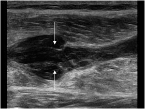
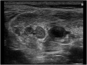
Cystic structure between the medial gastrocnemius and the soleus muscle.

Clinical information

Felt a sudden snap in the calf during kickboxing four weeks ago. There is not much clinical improvement and there is still a painful swelling of the calf.
Ultrasound Images & Clips

50 year old male, Felt a sudden snap in the calf during kickboxing four weeks ago. There is not much clinical improvement and there is still a painful swelling of the calf.
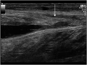
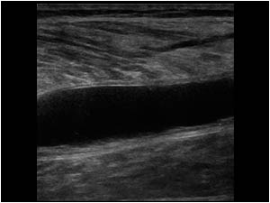
Longitudinal image of the calf with an anechoic structure between the medial gastrocnemius and the soleus muscle.
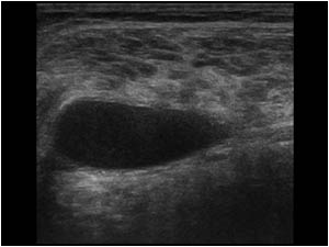
Transverse image of the same anechoic structure between the medial gastrocnemius an the soleus muscle.
The differential diagnosis of a fluid collection between the medial gastrocnemius and the soleus muscle include a hematoma caused by a gastrocnemius mu
Transverse image of the same anechoic structure between the medial gastrocnemius an the soleus muscle. The differential diagnosis of a fluid collection between the medial gastrocnemius and the soleus muscle include a hematoma caused by a gastrocnemius mu
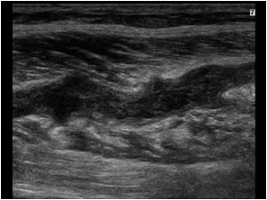
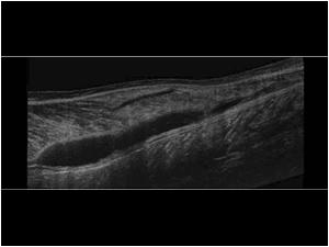
The extended field of view is very helpful to visualize the entire length of a lesion.
The lesion was over 13 cm long and had no relation with the knee space
The extended field of view is very helpful to visualize the entire length of a lesion. The lesion was over 13 cm long and had no relation with the knee space
This longitudinal image however shows a rupture with slight retraction of the stump(arrow) surrounded by a hematoma at the distal end of the medial gastrocnemius muscle.
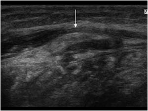
This transverse image shows a rupture with slight retraction of the stump(arrow) surrounded by a hematoma at the distal end of the medial gastrocnemius muscle.
In an uncomplicated case of a medial gastrocnemius muscle rupture or so called tennis leg, the
This transverse image shows a rupture with slight retraction of the stump(arrow) surrounded by a hematoma at the distal end of the medial gastrocnemius muscle. In an uncomplicated case of a medial gastrocnemius muscle rupture or so called tennis leg, the
Longitudinal image of the medial gastrocnemius muscle with a non compressible hypoechoic tubular structure.
Longitudinal image of the same tubular structure in the medial gastrocnemius muscle slightly more proximal.
There is a local widening of the  structure. The arrows point to the immobile valves in this thrombus filled vein.
Longitudinal image of the same tubular structure in the medial gastrocnemius muscle slightly more proximal. There is a local widening of the structure. The arrows point to the immobile valves in this thrombus filled vein.
Transverse image of the medial gastrocnemius muscle with non compressible thrombus filled veins.
The thrombosis of the gastrocnemius veins were a complication of the gastrocnemius rupture and could explain the impaired recovery.
In the case of venous th
Transverse image of the medial gastrocnemius muscle with non compressible thrombus filled veins. The thrombosis of the gastrocnemius veins were a complication of the gastrocnemius rupture and could explain the impaired recovery. In the case of venous th
Transverse image of the popliteal vessels. The popliteal vein is the more superficial structure. The artery is anechoic. But the vein shows an echogenic intraluminal structure (arrow).
Longitudinal image of the popliteal vessels. The arrow points to a thrombus mass extending into the popliteal vein.

Conclusion

Because of the extent of the thrombosis, the  patient was treated with anticoagulant therapy.

The combination of a deep venous thrombosis and a medial gastrocnemius rupture was first described in 1994.        

References            
Slawski DP. Deep venous thrombosis complicating rupture of the medial head of the gastrocnemius muscle. J Orthop Trauma. 1994;8(3):263-4.

Details

  • Sex: Male
  • Age: 50

Created with

  • Fujifilm Ultrasound System