March 2008

Poor visualisation of the right kidney with an echogenic rim around the kidney

Clinical information

Pain in the right upper abdomen
Ultrasound Images & Clips

48 year old female, Pain in the right upper abdomen

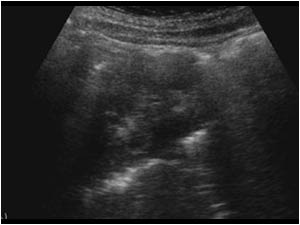
Longitudinal image of the right kidney with an echogenic rim around the kidney. The kidney is difficult to visualize.
Longitudinal image of the right kidney with highly reflective structures at the lower pole
Longitudinal image of the left kidney without any abnormality
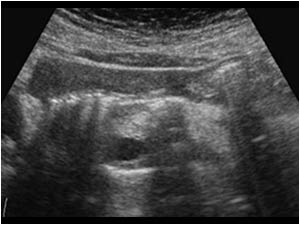
Transverse image of the pancreas. The head and a part of the body of the pancreas are obscured by the same echogenic structures.
Transverse image of the pancreas with the same echogenic structures that are clearly between the dorsal stomach wall and the pancreas and can therefore not be caused by air in the stomach.
What do you think causes the echogenic structures?
Transverse image of the pancreas with the same echogenic structures that are clearly between the dorsal stomach wall and the pancreas and can therefore not be caused by air in the stomach. What do you think causes the echogenic structures?
Patient also has gallstones.
Does this give you a clue to what could have happened?
Patient also has gallstones. Does this give you a clue to what could have happened?
A plain film of the abdomen makes clear why we had problems in examining the right kidney and pancreas. The image shows a massive amount of air surrounding the kidney consistent with a pneumoretroperitoneum.

Conclusion

The gallstones were the clue to the problems. A day before the ultrasound examination an attempt was made to perform an ERCP that had caused a duodenal perforation which caused the pneumoretroperitoneum.

References            
Nürnberg D, Mauch M, Spengler J, Holle A, Pannwitz H, Seitz KSonographical diagnosis of pneumoretroperitoneum as a result of retroperitoneal perforation]
Ultraschall Med. 2007 Dec;28(6):612-21.

Details

  • Sex: Female
  • Age: 48

Created with

  • Fujifilm Ultrasound System