November 2009

Abnormal appendix

Clinical information

Chronic pain in the right lower abdomen
Ultrasound Images & Clips

71 year old male, Chronic pain in the right lower abdomen

Longitudinal image of the appendix with a thickened distal end
Longitudinal image of the appendix. There is some vascularity within the thickened distal appendix.
Transverse image of the base of the appendix showing a normal oval appendix
Transverse image of the appendix a bit more distally. There is slight thickening of the appendix.
Transverse image of the appendix more distally. There is a thickened wall of the appendix.
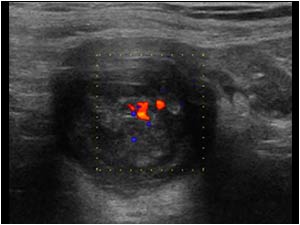
Transverse image of the tip of the appendix. There is a vascularized structure within the thickened appendix.
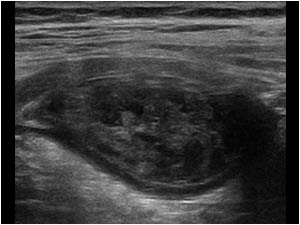
Longitudinal high resolution image of the appendix showing an intraluminal
Longitudinal high resolution image of the appendix showing an intraluminal "mass" in the thickened appendix
CT image of the thickened appendix with similar findings. There is minimal fat stranding around the thickened appendix.
Sagittal CT image of the appendix with similar findings.

Conclusion

Our initial differential diagnosis included a cecal carcinoma or a carcinoid of the appendix. The patient was operated. The diagnosis was a cystadenoma of the appendix with hyperplasic changes. The hyperplasic changes resulted in the vascularity within the thickened appendix mimicking a tumor. A cystadenoma was formerly called a mucocele of the appendix. A mucocele of the appendix is an infrequent event, representing 0.3%-0.7% of appendiceal pathology and 8% of appendiceal tumors. It is characterized by a located or diffuse distension of the appendix with a mucus-filled lumen. Spilling of the mucous content can cause pseudomyxoma peritonei. The diagnosis is often made by surgical intervention.
In case of an appendiceal mass, one must be aware of the possibility of one of these diagnoses.
In case of a cystadenoma of the appendix, follow-up is recommended, because sometimes they are associated with colorectal neoplasms and recurrence as pseudomyxoma peritonei.

References            
Kim SH, Lim HK, Lee WJ, Lim JH, Byun JY. Mucocele of the appendix: ultrasonographic and CT findings. Abdom Imaging. 1998 May-Jun;23(3):292-6.

Details

  • Sex: Male
  • Age: 71

Created with

  • Fujifilm Ultrasound System