For more than a year the patient had a painful right lateral elbow. The patient had pain (VAS 3) but experienced no functional limitations. She was referred by a general practitioner.
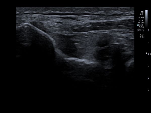
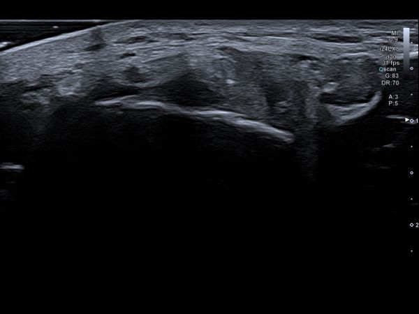
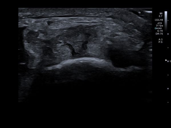
The transducer was placed both longitudinal (long-axis) and transverse (short-axis) over lateral epicondyle. The common extensor tendon was followed to distal passed the point of the radial neck. B-mode images where made both statically and dynamically. Additionally power Doppler measurements where made. A linear 24 Mhz and a Hockeystick 22 Mhz transducer were used. The Hockeystick was additionally chosen to visualize the most hypoechoic zone in the tendon. Images were systematically analyzed by using the SonoSkills pathology checklist.
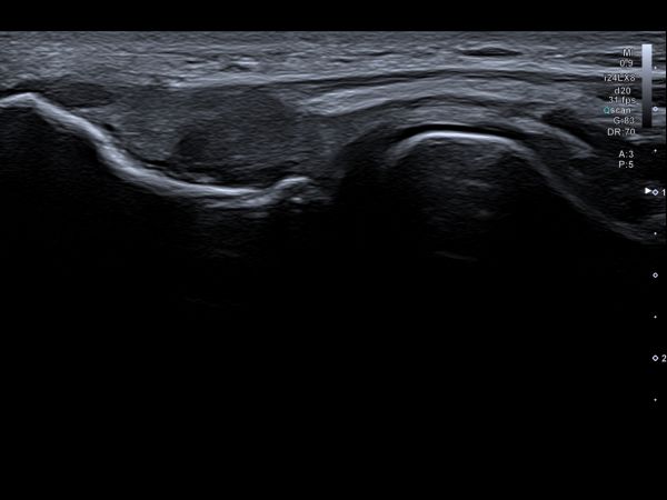
SHAPE: no changes in bone, joint and tendon morphology can be observed.
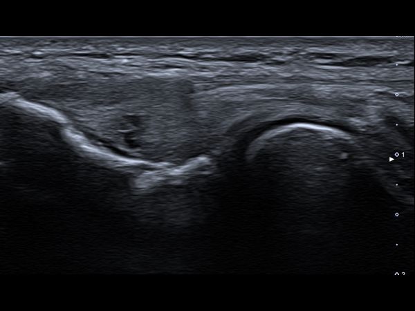
ECHOGENICITY: The common extensor tendons is much more hypo-echoic; zones in the deep and mid portion tendon seems to be anechoic.
CONTINUITY: There seem to be 2 or 3 partial tears in the deep and mid portion common extensor tendon; fibres appear to have been torn as no fibreconnection can be seen at those spots.
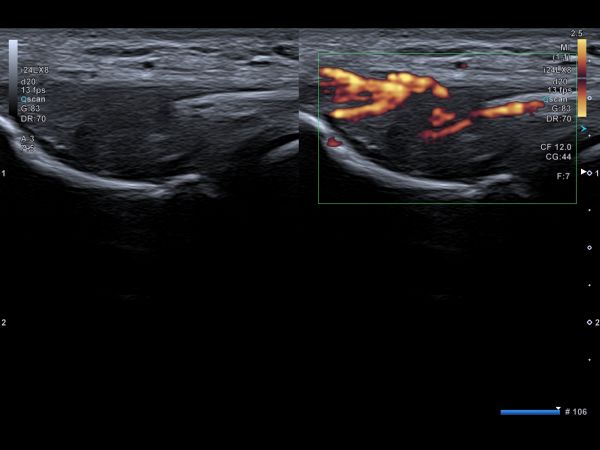
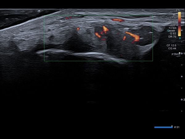
DOPPLER: power Doppler shows a grade 3 (range 0-3) neovascularization.
FUNCTIONAL: No functional limitations could be observed during ultrasound guided functional assessment.

Based on the ultrasound findings and SonoSkills pathology checklist analysis I concluded:
Moderate to severe tendinopathy of the common extensor tendon.
Moderate to severe partial tear in the deep and mid portion common extensor tendon.
Potential injury of the radial collateral ligament injury.
Grade 3 neovascularization


Marc is founder of, and trainer at, SonoSkills. SonoSkills is an organization dedicated to MSK ultrasound education. He's also an MSK Sonographer at the Laurentius Hospital in Roermond, the Netherlands.
More Cases from Marc Schmitz

