September 2006

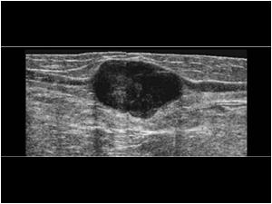
Hypoechoic mass in continuation with the greater saphenous vein.

Clinical information

Pregnant female 36 years old. Tumor in the upperleg since 2 years but increasing in size during the last few months.
Ultrasound Images & Clips

Pregnant female 36 years old, Tumor in the upperleg since 2 years but increasing in size during the last few months.

Longitudinal image of the mass in continuation with the greater saphenous vein. The mass is hypoechoic but solid, has a slightly lobulated aspect, smooth margins and shows no signs of infiltration.
Longitudinal image of the distal end of the mass and the greater saphenous vein.
Longitudinal image of the proximal end of the mass and the greater saphenous vein.
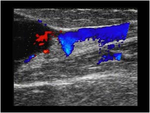
Longitudinal color doppler image of the distal end of the mass and the greater saphenous vein. The vein is still patent.
Longitudinal color doppler image of the proximal end of the mass and the greater saphenous vein. The vein is still patent.
Longitudinal color doppler image of the mass The mass is vascularized.
Cineloop shows flow in the mass with eflow
This is a longitudinal image of a small vascularized hypoechoic tumor in the cephalic vein of another pregnant female age 31. (The lumen of the vein is echogenic caused by slow bloodflow caused by external compression to demonstrate the lumen of the vesse
The mass is also vascularized.
There is flow in and along the mass. Surgical resection proved this mass to be a benign fibrous tumor.

Conclusion

What would you do in this case? Would you wait because the patient is pregnant and the mass is there for 2 years and has smooth margins?

The irregular vascularization and a previous case of a malignant tumor in continuation with the greater saphenous vein (case 7.4.4 slide 1) made us decide to perform an ultrasound guided biopsy.
Biopsy proved the lesion to be a malignant leiomyosarcoma.

Leiomyosarcomas are usually hypoechoic with smooth margins. They are uncommon tumors arising of smooth muscle cells. They acount for about 7 percent of all sarcomas. Uterus stomach and intestines are much more common sites than peripheral vessels.

The patient was operated and the mass proved to be a high grade leiomyosarcoma of the wall of the greater saphenous vein.

Not all tumors in peripheral veins are malignant.

References
Le Minh T,Cazaban D Great saphenous vein leiomyosarcoma: a rare malignant tumor of the extremity--two case reports. Ann Vasc Surg. 2004 Mar;18(2):234-6
Kreft B, Flacke S, Diagnostic imaging of vascular leiomyosarcomas Rofo. 2004 Feb;176(2):183-90.
Louail B, Vautier-Rodary R, Value of imaging in early diagnosis of peripheral vein tumors J Radiol. 1998 Nov;79(11):1387-91

Details

  • Sex: Female
  • Age: 36

Created with

  • Fujifilm Ultrasound System