Transperineal Biopsy of a Paediatric Prostate

Systematic biopsy of a 15-year-old male’s prostate gland

Clinical information

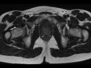
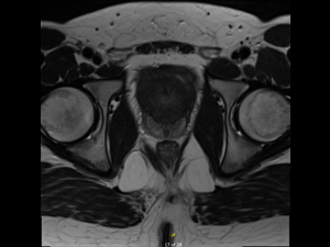
A 15-year-old male had been under the care of the paediatric urology team for long-term bladder outflow obstruction secondary to prostate enlargement for several years. He had had a previous transrectal ultrasound (TRUS) biopsy some years prior which had demonstrated no features of malignancy, but serial MRI scans had revealed progressive enlargement of the gland, such that there was sufficient concern to warrant a repeat biopsy. (See MRI images below)

Brief description

This coincided with the development of the transperineal biopsy service at the patient’s local university hospital, with the obvious benefits of a more extensive, systematic biopsy and the reduced risk of associated post-biopsy infection. The patient was therefore planned for a systematic transperineal prostate biopsy under general anesthesia, utilising the FujiFilm Arietta 65 ultrasound system with biplanar transperineal ultrasound probe C41L47RP.
Ultrasound Images & Clips

Axial T2 image through the prostate from 2015 demonstrates a significantly enlarged prostate returning homogeneous low T2 signal, a catheter in the urethra and a small midline prostatic cyst. The zonal anatomy is not well delineated.
Axial T2 image through the prostate from 2021 demonstrates further interval enlargement of the prostate demonstrating the typical high T2 signal peripheral zone and the bland low T2 signal transitional zone encroaching on the bladder neck.
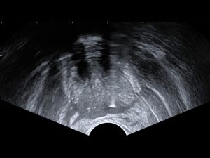
Transrectal ultrasound image in axial view of the prostate gland demonstrating biopsy of L posterior sector, peripheral zone
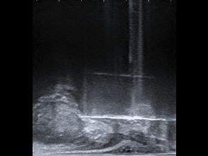
Transrectal ultrasound image of prostate gland in sagittal plane with biopsy needle targeting L mid sector, pushing through pseudocapsule to take core
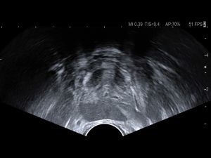
Axial view of the prostate gland at midline with no needle inserted. Note surrounding perimeter of fibromuscular tissue (prostate capsule) and urethra location.
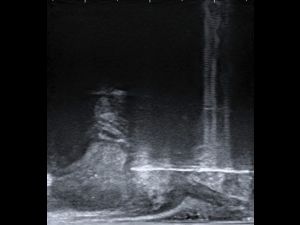
Transrectal ultrasound image of prostate gland in sagittal plane with biopsy needle targeting R posterior sector, peripheral zone with a lateral target (more medial).
Transrectal ultrasound image of prostate gland in sagittal plane with biopsy needle targeting R posterior sector, peripheral zone with a lateral target

Conclusion

A four-quadrant systematic prostate biopsy was performed, obtaining four 20mm 18-gauge biopsies from each quadrant, totalling 16 cores in total. The histopathology demonstrated features of chronic inflammation on a background of features consistent with a rhabdomyoma. There were no features to suggest malignancy and as such the patient will be kept under surveillance, with no surgical management needed at this juncture.